22
Mar
The navicular bone is the highest point and the keystone of the arch as it sits between the talus and cuneiforms. Redness and pain on palpation of the foot.
Navicular bone pain when i walk. PRP and stem cells are effective nonsurgical natural treatment options for Navicular Bone pain. The Navicular Bone is a small C shaped bone located on the inside portion of the midfoot. It provides important support of the foot and arch during movement.
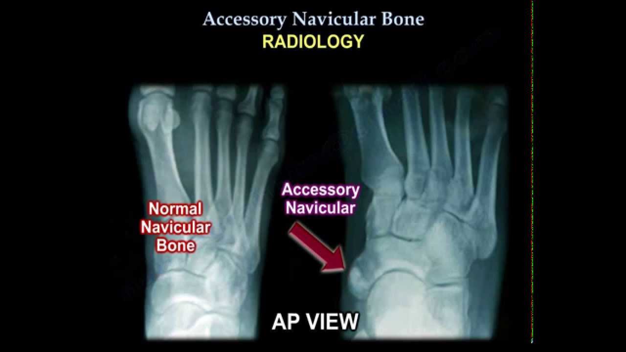
Fracture and arthritis are common causes of pain. Less common but other important causes of. The symptom most associated with accessory navicular is pain in the instep which can be aggravated by walking 2If the accessory navicular is to become painful it will typically happen during the teen years 1 2If the accessory navicular does not cause pain no nonsurgical or surgical treatment is needed 2Physicians identify the problem when the patient reports pain.
The most common symptom of navicular stress fractures is a persistent achiness in the arch or midsection of the foot that becomes worse with exercise or from prolonged standing. Sometimes pain can radiate along the inside edge of the foot temporarily resolving with rest and recurring when activity is resumed. The pain may be worse after athletic activity or just normal walking and walking itself may become painful.
This pain may become constant but it will tend to improve with continued rest. Depending on the size of the bump it may rub against shoes causing pain while walking or causing pain if the bump is hit by something. People who have an accessory navicular often are unaware of the condition if it causes no problems.
However some people with this extra bone develop a painful condition known as accessory navicular syndrome when the bone andor posterior tibial tendon are aggravated. This can result from any of the following. Trauma as in a foot or ankle sprain.
Navicular injuries are really tough to diagnose without a bone scan or MRI or in my case both. The bone is in the mid-foot and part of it can be touched on the top of the foot and another part on the inside near the arch. People describe varying symptoms for example mine never hurt when I pushed on the bone but most peoples do.
Stress fractures can occur from repeated stress. Athletes commonly fracture the navicular bone while kicking twisting or sprinting. Pain and change in how you walk are common with fractures.
Among track athletes navicular stress fractures are one of the most common causes of stress fractures. In some instances the best treatment is to remove the accessory navicular bone. This can end pain when walking though several weeks recovery time and some physical therapy could be required after the surgery.
Pain around cuneiform metatarsal and navicular bone. I felt some discomfort around the cuneiform bone on one of my feet. In terms of discomfort level it was low 2 or 3 out of ten i continued to walk around.
Yesterday the pain was slightly more frequent and only when I applie weight andwalked around. Broken foot navicular bone fracture The navicular is one of the bones of the foot. Difficulty walking constant foot pain pain in one foot recent ankle injury foot pain from overuse.
Symptoms that always occur with broken foot navicular bone fracture. Pain in one foot constant foot pain recent ankle injury. Pain with walking running or jumping.
Tenderness when pushing on the navicular bone. What does a navicular fracture feel like. Symptoms of a navicular stress fracture usually involve a dull aching pain in the ankle or at the middle or top of the foot.
In the early stages pain often occurs only with activity. In the later stages pain may be constant. Symptoms of a navicular stress fracture usually involve a dull aching pain in the ankle or at the middle or top of the foot.
In the early stages pain often occurs only with activity. In the later stages pain may be constant. Other common symptoms include.
Swelling or bruising over the middle part of the foot Limping. If people suffered a fracture of their navicular in a traumatic accident there are several symptoms that they might notice. Some of the most common problems include.
Swelling of the foot particularly in the middle. Pain that gets worse while walking or moving the foot. Redness and pain on palpation of the foot.
The most common causes of top of the foot pain. Top of the foot nerve compression. A swollen top of the foot can also occur due to stress across the top of the foot.
Lisfranc fracture or sprain. Sprained top of the foot. Top of the foot bone spur.
Hallux rigidus or dorsal 1st metatarsal joint spur. Dorsal foot compression syndrome. The navicular bone is a smaller rectangular shaped bone located immediately in front of the talus.
The talus bone is critical as it absorbs the forces of walking standing and running and transfers them to the other bones in the foot 1Running after talonavicular fusion is often impaired shortened by concentrated pain and tense overworked. Generally pain is worse with training and improves with rest. Often athletes can keep training as the pain is usually not as bad as other stress fractures.
As a result this leads to a delay in diagnosis. We suspect a navicular stress fracture if there is tenderness at the body of the navicular bone. Patient walks may also be evaluated.
X-rays are usually ordered to confirm the diagnosis. If there is ongoing pain or inflammation an MRI or other advanced imaging tests may be used to further evaluate the condition. Non-Surgical Approaches The goal of non-surgical treatment for accessory navicular syndrome is to relieve the symptoms.
The navicular bone is the highest point and the keystone of the arch as it sits between the talus and cuneiforms. The inner arch of the foot provides a great deal of the bodys weight bearing and shock absorption. The talus bears much of the weight and then transfers it forward through the navicular to the rest of the foot through the cuneiforms.
Previous post
Naxdom 250 side effects